

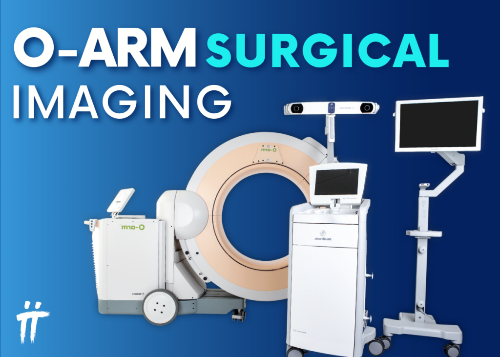
The next-generation O-ARM™ (intraoperative computed tomography) is a mobile imaging system that allows surgeons to visualize the patient’s changing anatomy in 2D and 3D with a 360-degree scan during surgery.
Precision is critical for success in spinal surgery. Accordingly, O-ARM™ provides surgeons with accurate, detailed, real-time imaging to help precisely align and place instruments and implants.
O-Arm Features
With the next-generation O-ARM™ imaging system, multidimensional images of patients’ spinal anatomy can be obtained in about 30 seconds during spinal surgery.
The O-ARM™ imaging system has been developed for spinal surgeries involving disc diseases, spinal canal stenosis, spinal tumors, certain developmental spinal disorders, herniated discs, scoliosis (spinal curvature), or any procedure requiring 3D imaging.
Spinal screw surgeries are mostly performed using a three-arm X-ray device called fluoroscopy. However, while fluoroscopy provides 2D images, the images obtained through the O-ARM™ imaging system are 3D.
During spinal screw surgeries performed with the O-ARM™, the anatomical area where the screw should be placed can be determined with 1–2 mm precision. This prevents the screw from entering unwanted areas and increases the success rate of the surgery. Additionally, the risk of injury to blood vessels, nerves, spinal cord, and vital organs is reduced, allowing spinal surgery to be performed safely.
Advantages
The advantages of spinal screw surgeries performed using the O-ARM™ surgical imaging system include:
- The amount of radiation exposure to patients is significantly reduced.
- Surgeons receive real-time information during all stages of surgery, allowing simultaneous visualization of the screw’s position and angle within the bone.
- Smaller incisions lead to shorter hospital stays and recovery times, reduced risk of infection, and less bleeding.
- Minimizes the major risks associated with complex surgeries.
- Reduces the risk of nerve paralysis caused by potential spinal cord injuries.
How to Apply?
Thanks to its robotic positioning feature, the O-ARM™ can be quickly positioned around the operating table in the surgical environment, allowing simultaneous image capture. It provides high-quality, multidimensional imaging with lower radiation compared to standard CT devices.
In the operating room, the O-ARM™ forms a ring around the patient’s body. The ring can be opened and closed, eliminating the need to move the patient. The device rotates to capture 2D real-time fluoroscopic images and 3D scans. Additionally, the O-ARM™ includes a navigation component similar to a GPS system, allowing the surgeon to track and guide instruments inside the body in real time for more precise hardware placement.
With its advanced features exceeding expectations, the next-generation intraoperative 3D imaging system:
- Provides enhanced image quality for more challenging patient anatomies, such as the cervicothoracic junction and pelvis,
- Offers surgeons options for orthopedic trauma procedures with 15 cm x 20 cm or 15 cm x 40 cm axial views and multidimensional imaging,
- Provides surgeons with real-time images,
- Adapts to surgical procedure workflows,
- Moves smoothly within the surgical environment.
O-ARM™-assisted procedures can improve outcomes in spinal surgery by shortening patients’ recovery times and enhancing postoperative results.


Leave a Comment